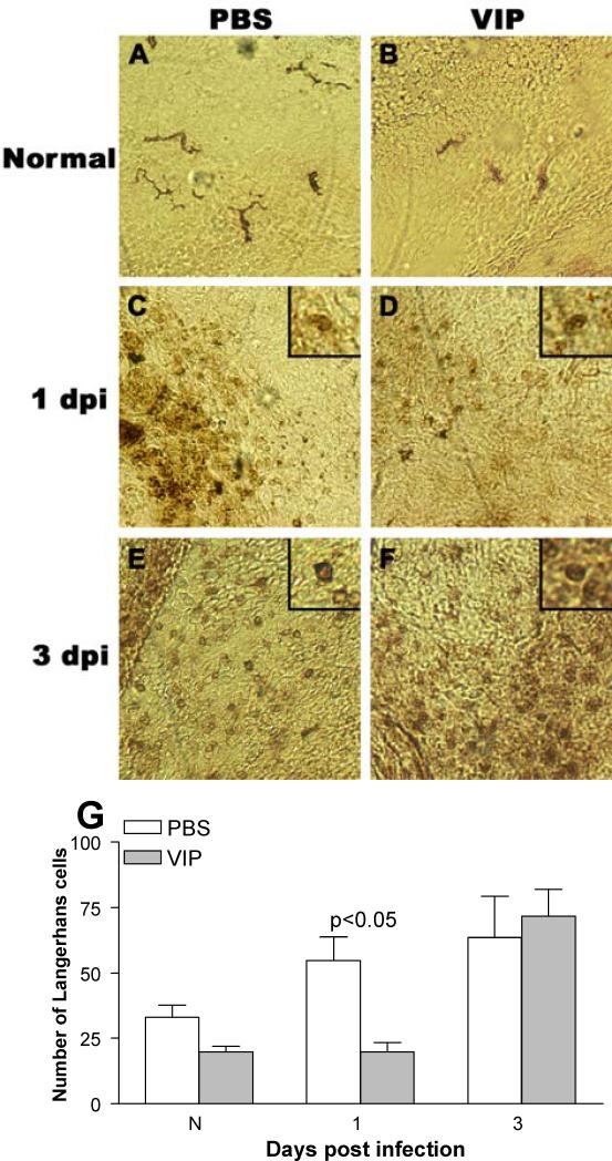
Figure 7.
Staining and quantitation of Langerhans cells. In the normal, uninfected eye (conjunctiva/peripheral cornea) dendritic-shaped ADPase positive Langerhans cells appeared similar in number after VIP (B) vs PBS (A). At 1 day p.i., rounded ADPase positive cells in the peripheral cornea appeared decreased after VIP (D) vs PBS (C) treatment. At 3 days p.i. cells qualitatively appeared similar after PBS (E) or VIP (F) treatment. Quantitation confirmed these findings (G). (A-F at 375 X magnification, field size = 270 μm2).

