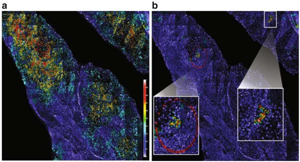
Fig. 7.
Automated detection of selected clonal populations of cells within a cancer biopsy tissue section. All nuclei (~150,000 in this example) are detected, and FISH probe signal counts are enumerated for each nucleus. FISH signal pattern for each cell is compared against its neighbor in order to define spatial association (or neighborhood). A mathematical model is then applied to determine clonal cell relationships. a Mapping cancer cells on a tissue section. A gain or loss of any one of three FISH markers indicates a cancer cell. This image shows the density of cancer cells (so defined) in neighborhoods as a color overlay. Red indicates high fraction of cancer cells, yellow indicates medium fraction of cancer cells, and blue indicates low to none (see scale bar). Most of the section is highlighted except for the surrounding normal stromal infiltrates. b Mapping clonal cells. The same image data were analyzed for concurrent gains of each of the three markers. The two clusters of cells, magnified within the white boxes, are cells harboring gain of all three markers

