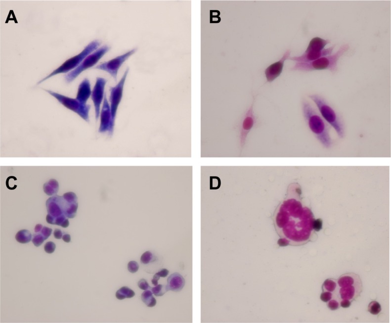
Figure 3.
Morphological features of LOVO cells after treatment for 48 hours by optical microscope (×400, Wright staining). (A) Controls, (B) 60 μg/mL magnetic nanoparticles containing Fe3O4, (C) 0.35 μmol/L gambogic acid, and (D) magnetic nanoparticles containing Fe3O4 and gambogic acid (0.35 μmol/L gambogic acid with 60 μg/mL magnetic nanoparticles containing Fe3O4).

