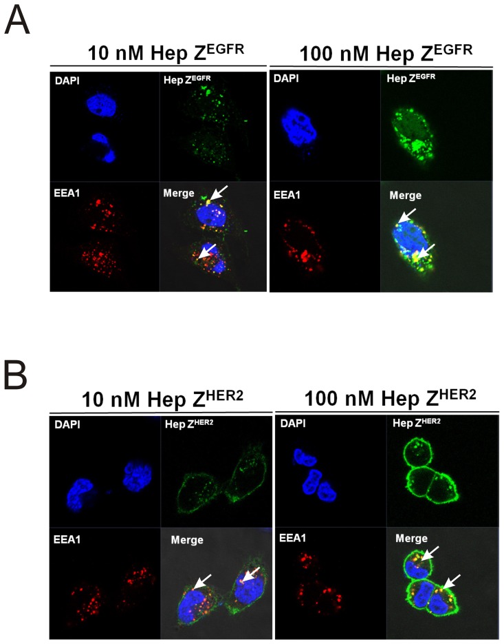
Figure 10. Co-localization of EEA1 and heptameric targeting ligands.
(A) Two different concentrations of the FITC-labeled heptameric ZEGFR targeting ligands were incubated with A431 cells for 2 h at 37°C. (B) FITC labeled heptameric ZHER2 targeting ligands at two concentrations were incubated with SK-OV3 cells for 2 h at 37°C. EEA1 proteins were detected by Alexa 555-conjugated secondary antibody. Top left panels: cell nuclei stained with DAPI (blue); Top right panels: FITC labeled heptamer (green); bottom left panels: EEA1 antibody (red); bottom right panels: merged image of the three stainings.

