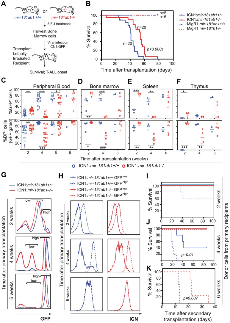
Figure 3. Loss of mir-181ab1 delays T-ALL and inhibits T-ALL development induced by low levels of ICN1.
(A) Schematic of experimental design. (B) Kaplan-Meier survival curves show the effect of loss of mir-181ab1 on the percentages of mice surviving at different time points after T-ALL induction with ICN1 (p<0.0001, n = 20 mice/experimental group, a representative plot of 4 independent experiments is shown). (C–F) Effects of loss of mir-181ab1 on the percentage of total ICN1-infected cells (all GFP+ cells) and the percent of ICN1-infected DP leukemia cells (GFP+DP cells) in (C) peripheral blood, (D) bone marrow, (E) spleen and (F) thymus of T-ALL mice (*, p<0.05; **, p<0.01; ***, p<0.001;). (G) Mice transplanted with ICN1:181ab1+/+ or ICN1:181ab1−/− BM cells have distinct profiles of GFP+ cells at 2, 4 and 6 weeks after transplantation. Gates that define GFPhigh and GFPlow cells are indicated. (H) Relative levels of ICN1 expression in GFPhigh and GFPlow cells were determined by intracellular staining of ICN1 and FACS analyses. (I–K) The Kaplan-Meier survival curves of the secondary transplantation analyses. ICN1-infected DP cells (GFP+ DP cells) were sorted from primary recipients at (I) 2, (J) 4 and (K) 6 weeks after transplantation and transferred to each of five secondary recipients. Secondary recipients were analyzed for survival and development of DP leukemia cells in the peripheral blood to determine the leukemogenic potentials of GFPhigh and GFPlow cells from primary recipients transplanted with ICN1:181ab1+/+ and ICN1:181ab1−/− BM cells.

