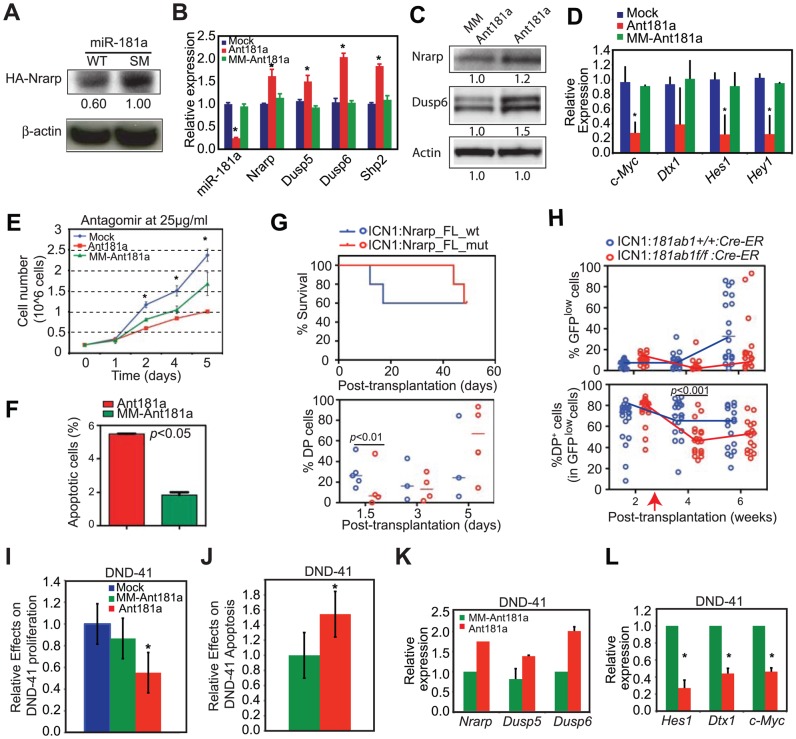
Figure 8. Targets of miR-181a in T-ALL cells and the effects of miR-181a inhibition in T-ALL cells.
(A) Western blot analyses were used to determine the levels of HA-tagged Nrarp protein in T6E cell lines expressing wild-type miR-181a or miR-181aSM (normalized to β-actin loading control). (B–D) Levels of miR-181a target expression determined by (B) qPCR or (C) western blot analyses and (D) Notch1 target expression in mock-treated T6E cells or cells treated with 10 µg/ml antagomir against miR-181a (Ant181a) or mismatch control (MM-Ant181a) for 48 hours (mean ± SD, n = 3, *p<0.05). (E and F) Effects of Ant181a and MM-Ant181a on T6E leukemia cell (E) proliferation and (F) apoptosis (mean ± SD, n = 3, *, p<0.05). (G) Kaplan-Meier survival curves and the percentages of ICN1-infected DP cells show the effects of Nrarp target expression on ICN1-induced T-ALL development (5 mice/group). Sorted ICN1-BM cells from primary recipient mice were infected with the same titer of virus expressing either Nrarp-FLmut (miR-181a-insensitive) or Nrarp-FLw t (miR-181a-sensitive) and transplanted into lethally irradiated recipients (5 mice/group). (H) Effects of conditional deletion of mir-181ab1 on the T-ALL development at 2 weeks after leukemia induction (see also Figures S8A, B). Percentages of GFPlow and GFPlow DP cells in the peripheral blood of recipient mice transplanted with either ICN1:181ab1+/+/Cre-ER BM cells or ICN1:181a1b1f/f/Cre-ER BM cells were determined by FACS analyses (20 mice/group). Red arrow indicates the initiation of CreER-mediated mir-181ab1 deletion. (I and J) Effects of Ant181a and MM-Ant181a (25 µg/ml) on human T-ALL DND41 cell (I) proliferation and (J) apoptosis after 96 h in culture (mean ± SD, n = 4, *, p<0.05). (K and L) Levels of (K) miR-181a targets and (L) Notch1 target expression in DND41 cells treated with Ant181a (25 µg/ml) for 48 hours (mean ± SD, n = 3, *p<0.05).

