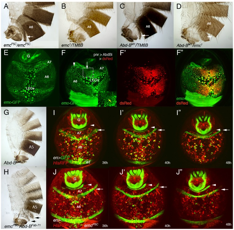
Figure 4. emc regulates extrusion of the male A7 histoblasts.
(A, B) emcP5C homozygous males show a small A7 segment (A), absent in emc− heterozygous conditions (B). (C) Males heterozygous for an Abd-B mutation present a small A7 [3], [4], and the trans-heterozygous combination Abd-BM1/emc1 enhances this phenotype (D). Arrows indicate the A7 segment. (E) Distribution of emc-GFP in the A7 and A6 segments of a ∼38 h APF male pupa. There is emc expression in LECs and histoblasts, with higher levels in A7 than in A6 histoblasts. We have measured the difference in signal intensity between nuclei of the two segments and found that the A7/A6 signal ratio is 1, 32±0,15 (n = 4). See also higher levels at the periphery of histoblast nests. (F–F″) UAS-DsRed/+; emc-GFP pnr-Gal4/UAS-Abd-BRNAi male pupa of about 36 h APF showing that in the central region of pnr expression (red in F′, F″), where Abd-B levels are reduced, emc-GFP levels are also reduced (arrow in F). Levels remain high where Abd-B has not been eliminated (arrowhead in F) but also in midline cells (also with high expression in anterior segments; yellow arrow in inset). (G) Abd-BFab7-1 homozygous adult male. The A6 disappears as it is transformed into the A7 [20]. (H) Abd-BFab7-1 emcP5C homozygous male: there are small A6 and A7 segments (arrows), showing the emcP5C mutation is epistatic over the Abd-BFab7-1 mutation. (I–I″) Snapshots from Video S12 in a ∼36–48 h APF en-Gal4 UAS-GFP/His2A-RFP male, showing the progressive disappearance of the A7 segment. Posterior compartments show en expression (in green), whereas nuclei are labeled in red. The arrow marks the A6p band of expression and the arrowhead the A8p cells. (J–J″) Snapshots from Video S13, taken at similar stages and with the same markers but in an emc mutant background (en-Gal4 UAS-GFP/His2A-RFP; emcP5/emcP5male). Note that, contrary to the previous panels (I–I″), the A6p en band does not move posteriorly, indicating that the A7 segment is not being extruded. Numbers indicate approximate hours APF.

