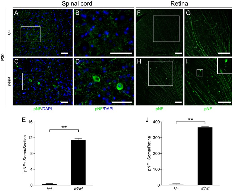
Figure 3. Phosphorylated neurofilament (pNF) accumulates in somas of spinal cord neurons and retinal ganglion cells in wl/wl mice.
Note the white boxes in A, C, F and H define areas that have been magnified in B, D, G and H, respectively. (A, B) At P30 pNF localized to only axons of lumbar motor neurons in wild type mice, but was not found in the somas of neurons. (C, D) At P30 in wl/wl mutant animals pNF was present in the axons of motor neurons, but also accumulated in the somas of some motor neurons. (E) Lumbar motor neuron sections stained with pNF antibody were used to count the number of soma in which pNF accumulated. There was a significant increase in the number of somas accumulating pNF in the motor neurons of wl/wl mice (12±0.4) compared to wild type mice (0.2±0.2). (F, G) In P30 wild type mice pNF labeled only ganglion cell axons in the retina, but in wl/wl mutants (H, I) pNF was also present in a subset of ganglion cell somas, especially in the peripheral region of the retina. In addition, focal swelling was observed in some axons. An enlargement of a focal swelling is shown in the upper right hand corner of I, which corresponds to the box in I. (J) Quantification of pNF+ somas confirms a massive increase in the number of somas accumulating pNF in wl/wl mice (364±7) compared to controls (2±0.5). **, P<0.01; Scale bar, 50 µm.

