
Figure 7. ATP8A2 expression and localization.
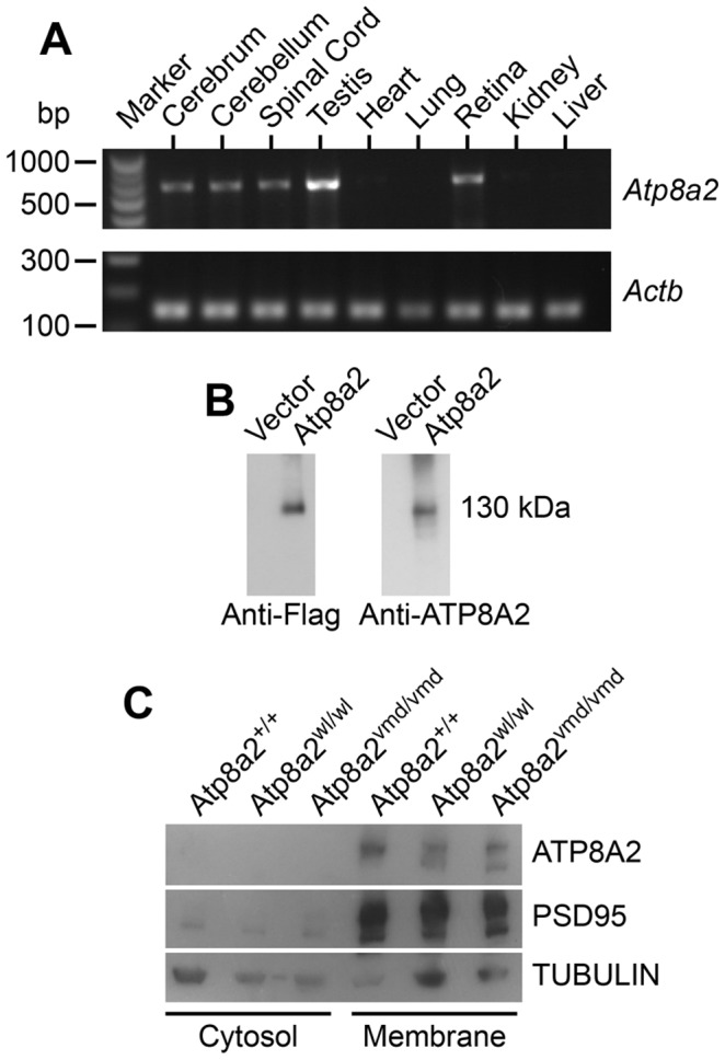
(A) Reverse transcript (RT)-PCR analysis showed that Atp8a2 is expressed in the cerebrum, cerebellum, spinal cord, testis and retina (Upper panel). The control for RT-PCR efficiency was β-actin (Actb; lower panel). (B) HEK293T cells were transfected either with mouse Atp8a2 cloned into pCMV6-AN-DDK vector, or empty vector in order to characterize the newly generated anti-ATP8A2 polyclonal rabbit antibody. Total protein (10 µg) isolated from these cells was used in western blot analysis with commercial anti-Flag antibody, or the anti-ATP8A2 antibody. Both Flag (left panel) and ATP8A2 antibodies (right panel) detected a protein of the expected 130 kDa in lysates from Atp8a2 cDNA transfected cells. (C) Total brain proteins were fractioned into membrane and cytosol fractions and subjected to SDS-PAGE and western blot analysis. ATP8A2 antibody detected the 130 kDa band only in the brain membrane fraction of wild type (+/+), wl (Atp8a2wl/wl) and vmd (Atp8a2vmd/vmd) mice. PSD95 was used as a membrane protein marker; TUBULIN was used as a marker for cytosolic proteins.