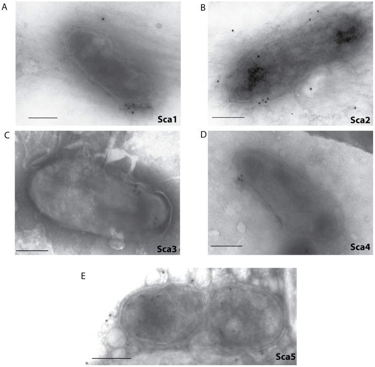
Figure 5. Immunogold electron microscopy of Sca expression on intact, negative stained R. typhi.
A suspension of rickettsiae was placed on grids and allowed to dry. Samples were labeled with antibodies against a Sca protein A) Sca1, B) Sca2, C) Sca3, D) Sca4 and E) Sca5 and negatively stained for contrast. Bar = 0.25 um.

