
Figure 6. dMi-2 does not promote chromatin decondensation by antagonizing histone H1 assembly.
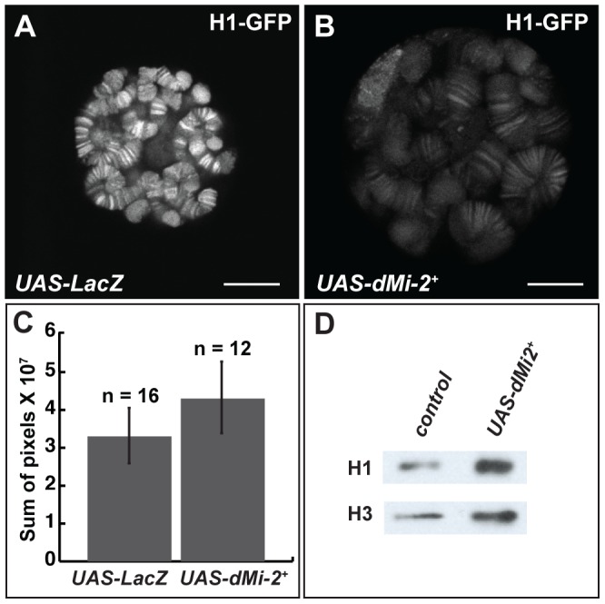
(A–B) Live analysis of salivary gland nuclei of late third-instar UAS-LacZ/+; ey-GAL4/+ (UAS-LacZ) control larvae (A) and UAS-dMi-2+ 3-3/+; UAS-dMi-2+ 15-1/ey-GAL4 (UAS-dMi-2+) larvae (B) expressing H1-GFP. Scale bars are 10 µm. (C) Quantification of H1-GFP fluorescence in larvae shown in A and B. The exposure times used to capture the images are identical; the number of glands analyzed is noted. (D) Protein blot showing the relative levels of histones H1 and H3 in chromatin extracted from late third-instar UAS-dMi-2+ 3-3/+; da-GAL4/+ (UAS-dMi-2+) and UAS-LacZ/+; da-GAL4/+ (UAS-LacZ) larvae raised at 29°C.