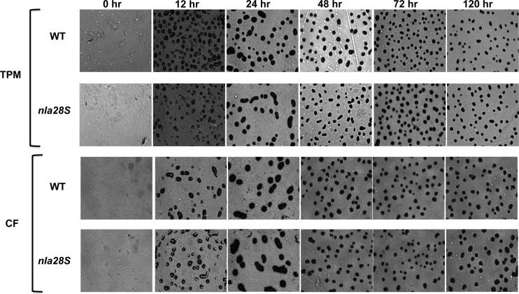
Fig 5.
Development phenotypes of wild-type (WT) and Δnla28S mutant cells. Wild-type and Δnla28S cells were spotted onto TPM agar plates (top two panels) or CF agar plates (bottom two panels), and the progress of fruiting-body development was monitored for 5 days using a Nikon Eclipse model E400 microscope at a total magnification of ×40. Photographs were taken at 0, 24, 48, 72, and 120 h poststarvation.

