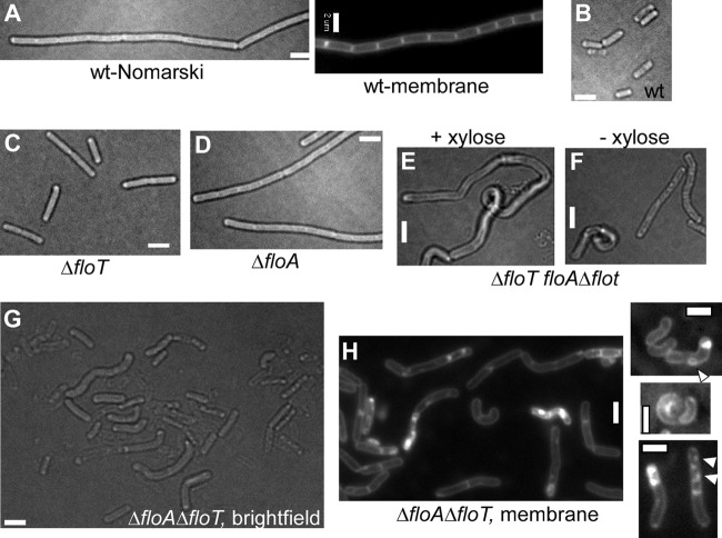
Fig 1.
Effects of deletions of flotillin-encoding genes on cell shape and cell division. (A and B) Chain of growing B. subtilis cells during vegetative growth (membrane stain FM4-64) (A) or in stationary phase (B). (C) ΔfloT cells during exponential growth. (D) ΔfloA cells during exponential growth. (E and F) Cells carrying a floT deletion and a floA truncation, in which the transcription of the yqfB gene is under the control of the xylose promoter, growing in the presence (E) or absence (F) of xylose. Note that there is no visual difference between the shape defects under the two conditions. (G) ΔfloT ΔfloA double-mutant cells during exponential growth. Note that lysed cells appear more transparent than living cells. (H) Membrane stain of exponentially growing ΔfloT ΔfloA double-mutant cells. The arrowheads on the right indicate membrane abnormalities. Bars, 2 μm.

