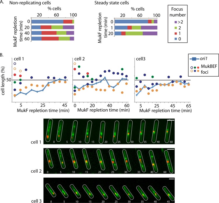
Fig 3.
Formation of MukB foci is independent of replication. (A) MukB focus appearance during MukF repletion is independent of replication or cell stage. The appearance and number of MukB foci were assessed before (0 min) and 20, 40, and 60 min after induction of MukF repletion (strain Ab174) in nonreplicating cells (left) and at 1 and 20 min after induction of MukF repletion in steady-state cells (strain Ab234). In the steady-state cells, transcription initiation was blocked at each time point by the addition of rifampin. The percentage of cells with 0, 1, 2, or >2 foci per cell are shown. n > 200. (B) MukB focus dynamics with respect to ori1 repositioning during MukF repletion in the absence of replication. Time-lapse analysis of MukB focus formation and ori1 positioning during MukF repletion in nonreplicating cells (strain Ab174). Time-lapse traces of ori1 (blue lines) as well as the position of MukB foci (circles) for three representative cells are shown. ori1, red; MukB, green. Cell outlines are in white. Images were taken every 5 min. Note that the 0-min time point in panel B is not equivalent to that in panel A, since there is a delay of ∼2 min between induction and imaging during the time-lapse experiments. Bars, 2 μm.

