Figure 1.

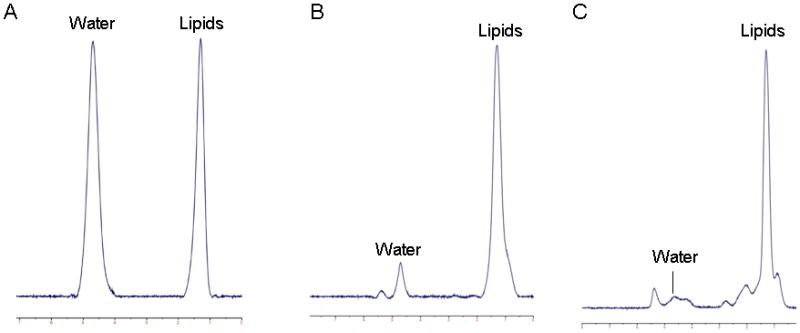
1H-magnetic resonance spectra of bone marrow in a subject with anorexia nervosa. Panel A: L4 vertebral body, 0.99 lipid to water ratio (LWR); Panel B: proximal femoral metaphysis, 5.7 LWR; Panel C: mid femoral diaphysis, 10.4 LWR.

1H-magnetic resonance spectra of bone marrow in a subject with anorexia nervosa. Panel A: L4 vertebral body, 0.99 lipid to water ratio (LWR); Panel B: proximal femoral metaphysis, 5.7 LWR; Panel C: mid femoral diaphysis, 10.4 LWR.