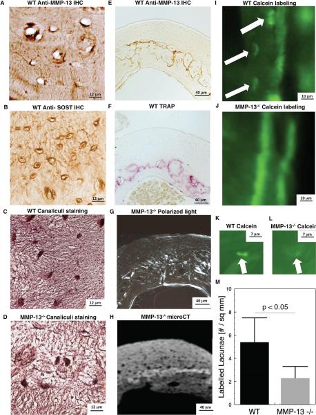
Figure 5. MMP-13 is required for normal osteocyte perilacunar matrix remodeling in mid-cortical bone.
IHC staining for MMP-13 (A) and SOST (B) show osteocyte lacunar and canalicular localization of both proteins in WT tibial cortical bone. Thionin staining shows that the normal canalicular organization of mid-cortical bone is disrupted by MMP-13 deficiency (C, D). MMP-13 localization in mid-cortical WT bone (E) overlaps with TRAP-staining in WT bone (F). Loss of MMP-13-mediated matrix remodeling by osteocytes causes collagen (G) and mineralization (H) defects in the same region of mid-cortical bone. Calcein-labeling of osteocyte lacunae near the periosteal surface (I) or in the mid-cortical region (K) of WT bone was less intense and less frequent in MMP-13−/− bone (J, L, M).

