
Figure 5.
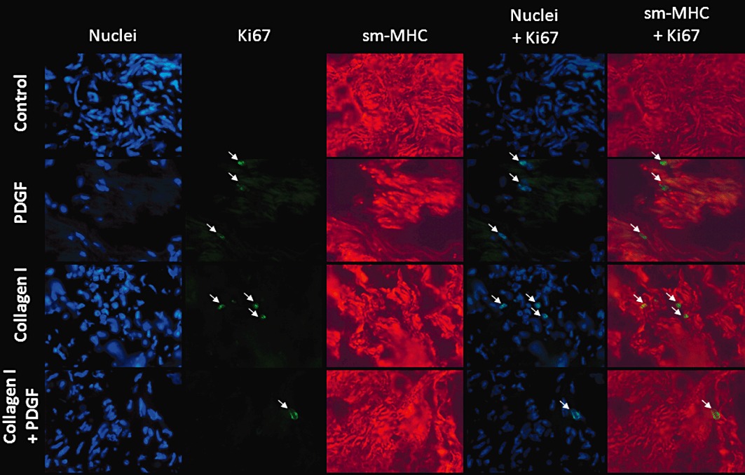
Immunofluorescent stainings of nuclei (Hoechst 33342), Ki67 (green), sm-MHC (red) in cryostat sections of HTSM strips treated without (control) or with collagen I (50 µg·mL−1) in the absence or presence of PDGF (10 ng·mL−1) for 4 days. Pictures were taken at a 400× magnification. Arrows indicate Ki67-positive nuclei.