Figure 8.

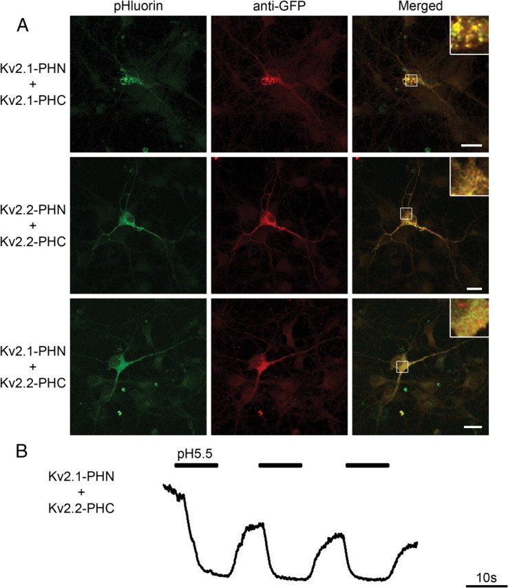
Bimolecular fluorescent complementation using Kv2.1 and Kv2.2 heteromers reveals distinct subcellular localization in vivo. A, Representative confocal images of hippocampal neurons transfected with either Kv2.1-PHN plus Kv2.1-PHC (top), Kv2.2-PHN plus Kv2.2-PHC (middle), or Kv2.1-PHN plus Kv2.2-PHC (bottom). Fluorescence from complemented pHluorin is shown on left (green). Neurons were fixed, permeabilized, and immunostained using antibodies against GFP to mark transfected cells (red). Merged image is shown on right. Regions of interest, denoted by white boxes, are shown at higher magnification below as inset at top right of merged image. Scale bars, 20 μm. B, Heteromeric Kv2.1–2.2 channel surface localization is revealed using acid quenching of surface fluorophore in hippocampal neurons. Representative traces from a hippocampal neuron transfected with Kv2.1-PHN plus Kv2.2-PHC demonstrate quenching of cell surface fluorophore at pH 5.5 (thick bar), which returns to baseline upon addition of solution at pH 7.4. Calibration: 10 s.
