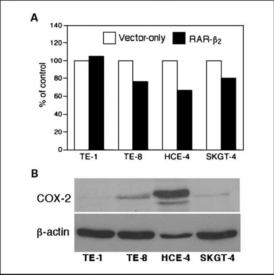
Fig. 4.
Reduced Ki67 positivity by transient transfection of RAR-β2 and association of COX-2 expression. A, cell proliferation assay. Esophageal cancer cell lines TE-1, TE-8, HCE-4, and SKGT-4 were transiently transfected with either pCMS/EGFP plus pRC/CMV vector or pCMS/EGFP plus pRC/CMV/ RAR-β2 for 2 d. The cells were then treated with 400 µg/mL G418 for additional 24 h and stained for Ki67 immunohistochemically. After that, more than 200 cells in 10 fields of 20× objective were counted for positive staining of GFP (green) as well as for positive or negative Ki67 (red) staining in these cells. The percentage of control of cell proliferation was calculated from the following equation: % control = NT/NV × 100, where NT and NV are the numbers of Ki67-positive cells in GFP-positive cells of RAR-β2–transfected and vector control cultures, respectively. B, Western blot analysis of COX-2 expression. The cells were grown in monolayer for 5 d and total cellular protein was extracted and subjected to Western blot analysis.

