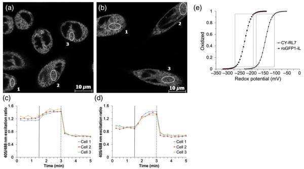
Figure 5.
Representative images of the roGFP1-iL sensor targeted to the ER of CHO cells. (c, d) Corresponding time-lapse responses of 405/488 nm ratio to sequential treatment with diamide (vertical solid line) and DTT (vertical dashed line) added to cells at concentrations of 1 and 10 mmol/L, respectively. The data are representative of six independent experiments using a minimum of three ROIs. (e) Relationship between the redox potentials in mV and the fractions of CY-RL7 (solid line) and roGFP1-iL (dashed line) in oxidized form. Boxes denote changes in probe fluorescence from 5 to 95% oxidation, which translates to an effective measuring range equal to a given probe midpoint potential ± 40 mV. ER, endoplasmic reticulum; CHO, Chinese hamster ovary; DTT, dithiothreitol

