Figure 2 .
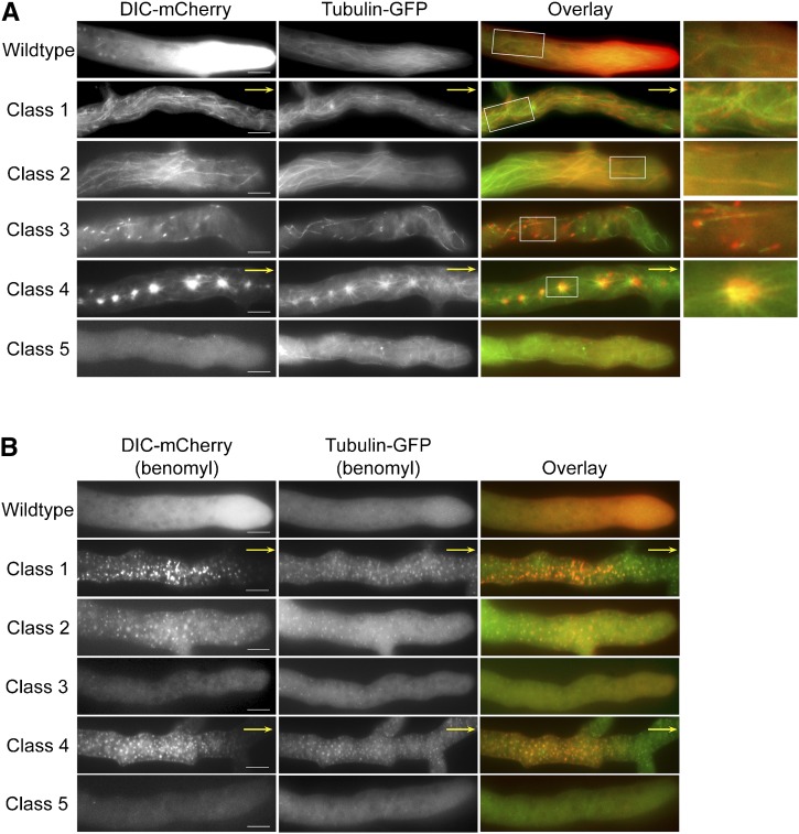
Localization of dynein and microtubules in wild-type and DHC mutant strains. (A) Epifluorescence images showing hyphal localization of DIC-mCherry (left panels) and β-tubulin-GFP (center panels) in wild-type and DHC mutant strains. Overlay images are shown in the right panels. Enlarged views of the boxed regions are shown on the far right. Yellow arrows indicate that the hyphal tips were located outside the frame. Dynein in wild-type, mutant class 1, 2, 3, and 4 strains was found along microtubule structures. Class 5 mutant strains did not show dynein colocalization with microtubules. Bars, 10 µm. (B) Dependence of dynein localization patterns on intact microtubule network. Treatment with benomyl depolymerizes microtubules and alters dynein localization in wild-type and DHC mutant strains expressing DIC-mCherry and β-tubulin-GFP. Dynein (red) is shown in left panels; microtubules (green) are shown in the center panels; overlay images are shown in the right panels. In wild-type and class 3 mutant strains, dynein localization to comet tails was disrupted immediately upon benomyl treatment. The cloud of dynein at the hyphal tips in wild-type strains disappeared after longer treatment times. Mutant class 1, class 2, and class 4 strains displayed punctate dynein signals that showed extensive overlap with microtubule remnants. Bars, 10 µm.

