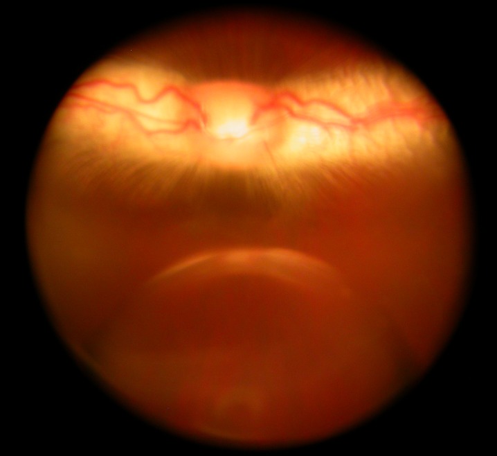
Figure 1. .
Fundus photograph demonstrates a bevacizumab droplet in a silicone oil-filled vitreous cavity, taken 18 hours after an intra-silicone oil injection. The droplet was floating in the vitreous cavity, and the optic nerve head and medullary ray show normal appearance. The droplet looked larger than its actual size because of eye refraction and camera focusing.

