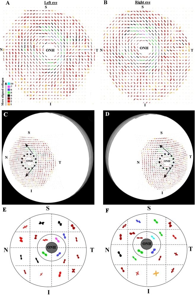
Figure 4. .
Preferential fiber orientation across non-glaucoma scleral pair. (A, B) Polar vector maps of preferential collagen fiber orientation across 15-mm diameter posterior scleral specimens (n1l and n1r) from a pair of non-glaucoma human eyes, sampled at 0.5-mm intervals, and viewed from the posterior side. The scaling of the vectors is related to the degree of anisotropy. To allow montage display, the larger plots (indicative of higher fiber anisotropy) have been scaled down by the factors indicated in the color key. Superior (S), inferior (I), nasal (N), and temporal (T) specimen positions are marked. The left eye has been mirror-flipped horizontally to enable direct comparison with the right eye. (C, D) Polar vector plots mapped onto 3-D posterior eye models. Dominant features of the fiber structure are indicated by arrows: note circumferential fibers ringing the optic nerve head (ONH) and the oblique splaying of meridional fibers from the ring toward the S-N and I-N directions. (E, F) Regional maps showing average polar plots within the four principal quadrants of the peripapillary sclera and 16 regions of the midposterior tissue.

