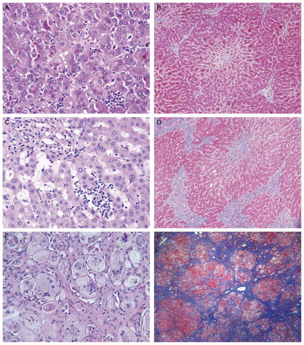
FIGURE 1.
Light microscopy findings in PFIC2 include varying lobular inflammation, portal inflammation, giant cell transformation, hepatocellular necrosis, hepatocellular swelling, portal fibrosis, and centrizonal/sinusoidal fibrosis. A and B, Explanted liver of patient 12 at the age of 17 months shows cholestasis, focal lobular inflammation, hepatocellular necrosis (including necrotic giant cells), prominent centrizonal fibrosis, and stage 2 portal fibrosis. Hematoxylin and eosin (H&E; × 400) and trichrome (× 100) stains. C and D, Explanted liver of patient 9 at the age of 7 years shows more prominent, diffuse lobular inflammation and areas of bridging fibrosis (shown) and nodule formation (not shown) with architectural distortion. H&E (× 400) and trichrome (× 100) stains. E and F, Explanted liver of patient 6 at the age of 16 months shows giant cells, hepatocyte swelling, and centrizonal/sinusoidal fibrosis, all prominent, and cirrhosis. Patchy intense lobular inflammation and pervasive bands of vascularized intralobular replacement fibrosis are seen. Central veins and portal-to-portal scars are not discernible; most of the fibrosis is pericellular. H&E (× 160) and trichrome (× 100) stains.

