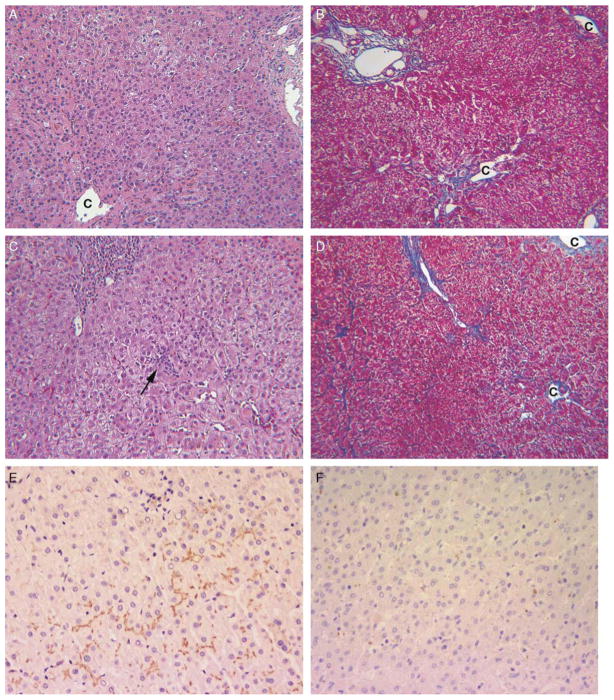
FIGURE 2.
Patient 7 had early-onset, intermittent pruritus and a long course (present age, 15 y), and his liver tissue showed intact canalicular BSEP immunostaining. Multiple liver biopsy specimens (A to D) showed stable liver disease with exacerbations of lobular cholestasis and hepatitis. A and B, Minimal cholestasis, occasional multinucleate giant hepatocytes, absent portal tract inflammation, and centrilobular fibrosis confined to zone 3 at the age of 13 months. C indicates central vein. Hematoxylin and eosin (H&E) and trichrome stains (each × 100). C and D, Mild portal and focal lobular inflammation (arrow) accompany lobular cholestasis and mild cell swelling at the age of 9 years. Fibrosis is mild. H&E and trichrome stains (each × 100) C indicates central vein. E, Immunostain for BSEP (hematoxylin counterstain) performed on liver biopsy tissue of patient 7 at the age of 9 years shows normal intensity and distribution of canalicular reactivity (× 200). F, In contrast, liver biopsy of patient 10 at the age of 21 months shows no canalicular reactivity. Immunostain for BSEP, hematoxylin counterstain (× 200).

