Fig 1.
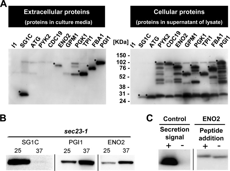
Detection of unconventional secretion of glycolytic enzymes. Anti-FLAG antibody was used for detection. (A) Secretion of glycolytic enzymes. (Left) Secreted proteins; (right) cellular proteins. I1, pULI1; SG1C, pULSG1C; ATG, pUL-ATG-EGFP; PYK2, pULGI2-PYK2; CDC19, pULGI2-CDC19; ENO2, pULGI2-ENO2; GPM1, pULGI2-GPM1; PGK1, pULGI2-PGK1; TPI1, pULGI2-TPI1; FBA1, pULGI2-FBA1; PGI1, pULGI2-PGI1. (B) The effect of inhibition of the conventional pathway on the secretion of the glycolytic enzymes. Secretion of recombinant proteins in sec23-1 strains at 25°C or 37°C is shown. (C) The effect of the N-terminal peptide (HA tag) on the secretion of enolase. Control (secretion signal +, secretion of EGFP-FLAG protein with conventional secretion signal sequence), pULSG1C; control (secretion signal -, secretion of EGFP-FLAG protein without secretion signal sequence), pUL-ATG-EGFP; ENO2 (peptide addition +, secretion of N-terminal HA peptide-tagged Eno2p-EGFP-FLAG), pULGI2-ATG-HA-ENO2; ENO2 (peptide addition -, secretion of Eno2p-EGFP-FLAG without peptide addition), pULGI2-ENO2. Similar results were obtained from 3 independent experiments. *, glycolytic enzymes conjugated to EGFP and FLAG. Additional bands are either nonspecific binding of antibody or degradation products of target proteins.

