
Fig 2.
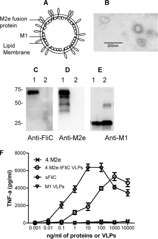
Characterization of 4.M2e-tFliC VLPs. (A) Diagram of influenza virus 4.M2e-tFliC VLPs. The membrane-anchored form of the 4.M2e-tFliC is inserted into the lipid bilayer of the envelope. (B) Negative-stain EM of VLP. (C to E) Characterization of VLPs. VLP samples were applied to SDS-polyacrylamide gels, followed by Western blotting. Lanes 1, 4.M2e-tFliC VLPs; lanes 2, M1-only VLPs. Protein bands were detected by mouse anti-FliC antibody (FliC-1) (C), mouse anti-M2e antibody (14C2) (D), and anti-M1 antibody (GA2B; Abcam) (E). (F) TLR5 agonist activity of flagellin. The mouse macrophage cell line RAW264.7, which expresses TLR2 and TLR4 without TLR5, was used to determine the bioactivity of 4.M2e-tFliC VLPs. Soluble recombinant FliC (sFliC) was used as a positive control; M1-only VLPs and recombinant 4.M2e were used as negative controls. TLR5-expressing RAW264.7 cells were prepared by transfection with the vector pUNO-hTLR5 expressing the human TLR5, as described previously (40). Data represent means ± standard deviations (SDs) from triplicate repeats.