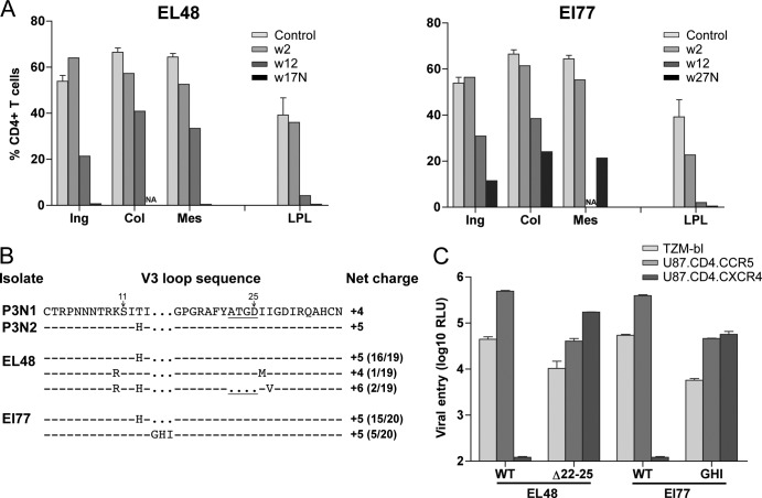
Fig 3.
Tissue CD4+ T cell frequency (A), V3 loop sequence (B), and coreceptor usage (C) of viral variants in macaques EL48 and EI77. (A) Percentages of CD4+ T cells in the inguinal (Ing), colonic (Col), and mesenteric (Mes) lymph nodes and lamina propria lymphocyte (LPL) from the jejunum during peak (w2) and chronic (w12) stage of infection and at time of necropsy (N) are reported. Baseline values generated from three uninfected macaques (control) are shown for reference. NA, not available. (B) V3 loop sequence comparison of representative SHIVSF162P3N clones (P3N1 and P3N2) and plasma viruses in macaques EL48 and EI77 at time of necropsy. Dots indicate gaps, and dashes stand for identity in sequences, with the net positive charge of the V3 region shown on the right. Positions 11 and 25 within the V3 loop are indicated by arrows, and the 4-amino-acid deletion is underlined. The numbers in parentheses represent the numbers of clones matching the indicated sequence per total number of clones sequenced. (C) Relative entry of pseudoviruses bearing EL48 and EI77 Envs into TZM-bl, U87.CD4.CCR5, and U87.CD4.CXCR4 indicator cells. RLU, relative light units. The data are means and standard deviations from triplicate wells and are representative of at least two independent experiments.

