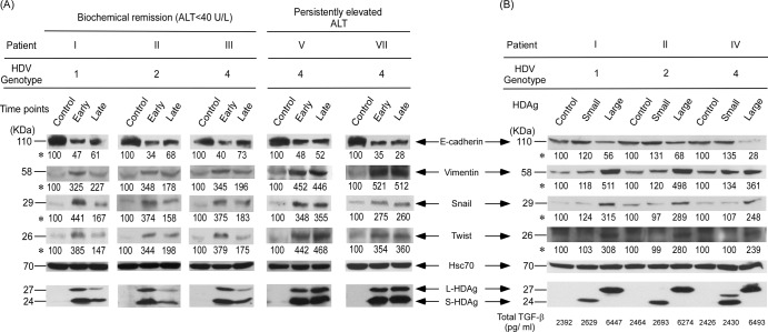
Fig 5.
Expression profiles of EMT markers determined by Western blotting. (A) EMT factor protein expression in Huh-7 cells transfected with the empty vector (pcDNA3.1) or the original or novel dominant HDV expression plasmid. (B) S-HDAg- or L-HDAg-expressing plasmids. Quantification of TGF-β1 was done with supernatants incubated under 2% FBS collected at the end of day 3. The basal level of TGF-β1 in culture medium without incubation with Huh-7 cells was 210 pg/ml. Intensities of bands shown for each EMT marker panel are indicated by asterisks. Statistical significance, P < 0.05 compared with the control, was determined by the two-tailed Student t test.

