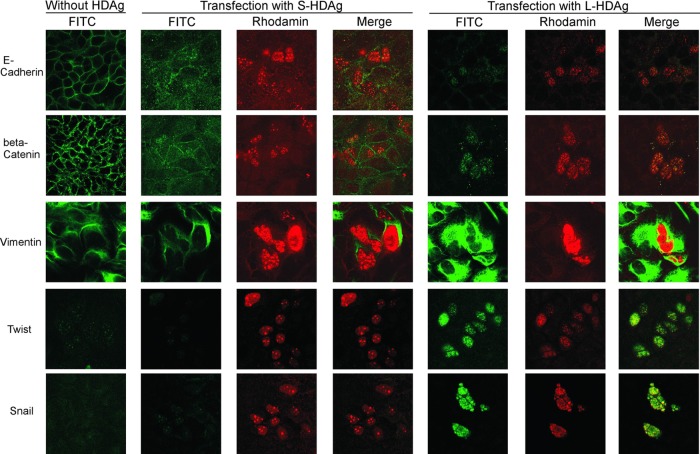
Fig 7.
Immunofluorescence staining of expressed EMT markers in Huh-7 cells transfected with large or small HDAg expression plasmids. S-HDAg and L-HDAg expression plasmids were transfected to Huh-7 cells (middle and right panels, respectively). The cells were doubly stained with human anti-HDV antibody and mouse or rabbit anti-EMT markers (E-cadherin, β-catenin, vimentin, twist, snail) after 3 days of transfection. Rhodamine-conjugated rabbit anti-human IgG, FITC-conjugated sheep anti-mouse IgG, or FITC-conjugated goat anti-rabbit IgG secondary antibodies were used to show the HDAg and EMT markers in red and green immunofluorescence stainings. Photographs (original magnification, ×630) were taken with a confocal fluorescence microscope (Axiovert 200M; Zeiss).

