Fig 1.
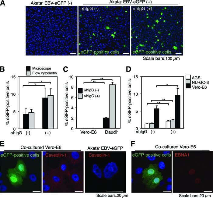
EBV is transmitted from BL cells to epithelial cells by cocultivation. (A) EBV transmission from BL cells to epithelial cells is facilitated by αhIgG treatment. Vero-E6 cells were cocultured with Akata− EBV-eGFP cells in the absence (middle) or presence (right) of αhIgG for 24 h. The infection of EBV-eGFP in Vero-E6 cells (green) was analyzed by a confocal laser scanning microscope. The left panel shows the result without cocultivation. The nucleus was counterstained with DAPI. Scale bars, 100 μm. (B) A summary of cell-to-cell contact-mediated EBV transmission. Vero-E6 cells were cocultured with Akata− EBV-eGFP cells in the absence or presence of αhIgG for 24 h. The percentages of EBV-eGFP-positive Vero-E6 cells were analyzed by a confocal laser scanning microscope (black bars) or flow cytometry (gray bars). The experiment was performed five times independently, and the averages and standard deviations are shown for each condition. *, P < 0.05 versus the respective control (Student's t test). (C) Infection of cell-free EBV-eGFP into Vero-E6 cells. Vero-E6 cells or Daudi− cells were incubated for 1 h at 37°C with culture supernatants derived from αhIgG-treated (gray bars) or untreated (black bars) Akata− EBV-eGFP cells. The culture supernatants were replaced with fresh medium, and the cells were further incubated for 48 h. The percentages of eGFP-positive cells were analyzed by flow cytometry. The experiment was performed three times independently. The averages and standard deviations are shown for each condition. **, P < 0.01 versus the respective control (Student's t test). (D) EBV-eGFP is transmitted to human gastric epithelial cells. AGS (white bars), NU-GC-3 (gray bars), or Vero-E6 (black bars) cells were cocultured with Akata− EBV-eGFP cells in the absence or presence of αhIgG for 24 h. The percentages of eGFP-positive epithelial cells were analyzed by flow cytometry. The experiment was performed three times independently, and the averages and standard deviations are shown for each condition. *, P < 0.05 versus the respective control. **, P < 0.01 versus the respective control (Student's t test). (E) EBV-eGFP is transmitted to Vero-E6 cells. Vero-E6 cells were cocultured with Akata− EBV-eGFP cells under the treatment of αhIgG for 24 h. The expression of an epithelium marker, caveolin-1, was analyzed by immunofluorescent staining. Caveolin-1 (red; middle) was expressed in eGFP-positive cells (green; left); BL-derived Akata− EBV-eGFP cells were caveolin-1 negative (right). The nucleus was counterstained with DAPI. Scale bars, 20 μm. (F) eGFP-positive Vero-E6 cells are infected with EBV-eGFP. Vero-E6 cells were cocultured with Akata− EBV-eGFP cells under treatment with αhIgG for 24 h. The expression of EBV-encoded nuclear antigen 1 (EBNA1) was analyzed by immunofluorescent staining. eGFP-positive Vero-E6 cells (green; left) were EBNA1 positive (red; right). The nucleus was counterstained with DAPI. Scale bars, 20 μm.

