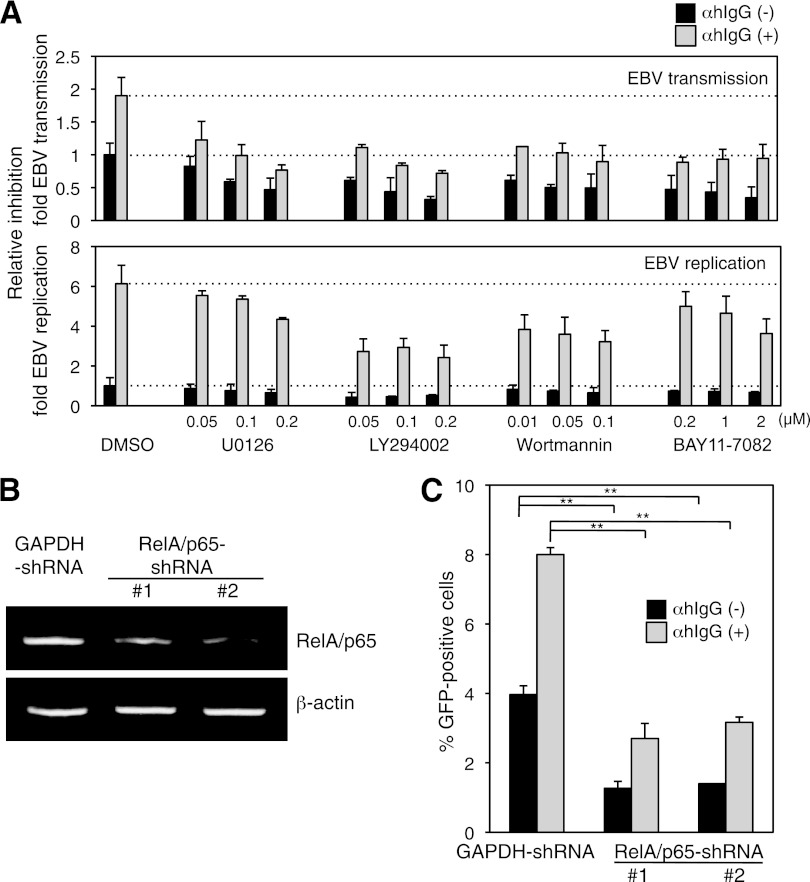
Fig 4.
Effect of the inhibitors of cell signaling pathways on cell-to-cell contact-mediated EBV transmission and replication. (A) The effect of the inhibitors on cell-to-cell contact-mediated EBV transmission (top) and EBV replication (bottom). Vero-E6 cells were cocultured with Akata− EBV-eGFP cells in the presence (gray bars) or absence (black bars) of αhIgG under treatment with DMSO, U0126, LY294002, wortmannin, or BAY11-7082 for 24 h. The percentages of eGFP-positive cells were analyzed by flow cytometry. The data were normalized to untreated and DMSO-treated cells (top). Cocultured Akata− EBV-eGFP cells were harvested, and the expression of gp350 was analyzed by flow cytometry. The data were normalized to αhIgG-untreated and DMSO-treated cells (bottom). The experiment was performed three times independently. The averages and standard deviations are shown for each condition. (B) RelA/p65 knockdown by shRNA in Vero-E6 cells. Total RNA was isolated from two Vero-E6 clones stably expressing shRNA against human NF-κB RelA/p65 (RelA/p65-shRNAs 1 and 2) and a control expressing shRNA against human glyceraldehyde-3-phosphate dehydrogenase (GAPDH-shRNA). Knockdown of RelA/p65 mRNA was analyzed by RT-PCR (top). As a control, the expression of β-actin mRNA is shown (bottom). (C) The effect of RelA/p65 knockdown on cell-to-cell contact-mediated EBV transmission. RelA/p65-shRNA or GAPDH-shRNA was cocultured with Akata− EBV-eGFP cells in the presence (gray bars) or absence (black bars) of αhIgG for 24 h. Viral transmission was assessed by flow cytometry. The experiment was performed three times independently. The averages and standard deviations are shown for each condition. **, P < 0.01 versus the respective control (Student's t test).

