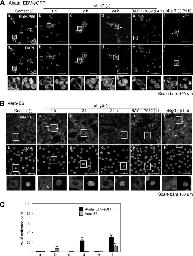
Fig 7.
Activation of the NF-κB pathway in cocultured cells. The nuclear translocation of RelA/p65 in cocultured Akata− EBV-eGFP (A) or Vero-E6 (B) cells. Vero-E6 cells were cocultured with Akata− EBV-eGFP cells in the presence (b to e) or absence (f) of αhIgG for various times. The translocation of RelA/p65 in Akata− EBV-eGFP (A) or Vero-E6 (B) cells was examined by immunofluorescent staining (top). The effect of BAY11-7082 treatment (2 μM) on the translocation of RelA/p65 is shown in image e. As a control, the status of RelA/p65 translocation in Akata− EBV-eGFP or Vero-E6 cells not treated with αhIgG is shown in image a. The nucleus is counterstained with DAPI (bottom). Insets show enlargements of the boxed areas. Scale bars, 100 μm. (C) The percentages of the cells that contain the translocation of RelA/p65 were analyzed by a confocal laser scanning microscope. The experiment was performed three times independently. The averages and standard deviations are shown for each condition. **, P < 0.01 versus the respective control (Student's t test).

