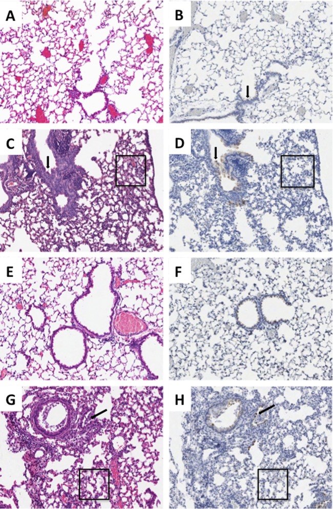
Fig 2.
Histopathology and influenza virus antigen distribution for selected viruses. Matched sections of day 5 postinfection lungs (original magnification, ×100). H&E stains are shown in the left column, and influenza virus A antigen immunohistochemistry is shown in the right column with the DAB end product counterstained with hematoxylin. (A and B) Infection with the AI virus, demonstrating the absence of histopathologic changes and influenza virus antigen except rare, multifocal labeling of the bronchial epithelium (arrow). (C and D) Infection with the 1918 virus, showing severe multifocal changes, including transmural necrotizing bronchitis (arrow) and multifocal moderate-to-severe alveolitis with multifocal pulmonary edema (box) and influenza virus antigen in bronchial epithelium (arrow) and alveolar epithelial cells. (E and F) Infection with the 1918AI-PB2 virus, showing a similar absence of histopathologic changes comparable to the AI virus, accompanied by slightly stronger labeling of bronchial epithelium. (G and H) Infection with 1918AI-PB2-K627, showing similar histopathology, including transmural necrotizing bronchitis (arrow) and multifocal moderate-to-severe alveolitis with multifocal pulmonary edema (box) and influenza virus antigen distribution in bronchial epithelium and alveolar epithelial cells similar in distribution to the parental 1918 virus.

