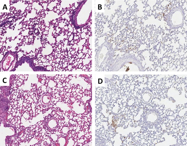
Fig 4.
Histopathology and neutrophil distribution for 1918 and 1918AI-HA viruses. Matched sections of day 5 postinfection lungs (original magnification, ×100). H&E stains are shown in the left column, and myeloperoxidase immunohistochemistry is shown in the right column with the DAB end product counterstained with hematoxylin. (A and B) 1918 virus-infected mice, showing severe multifocal changes and numerous myeloperoxidase-positive cells (neutrophils) prominently in the smallest airways, bronchioles, and alveoli. (C and D) 1918AI-HA virus-infected mice, showing similar histopathologic changes to 1918 and a comparable number of neutrophils in correlation with the histopathology.

