Fig 4.
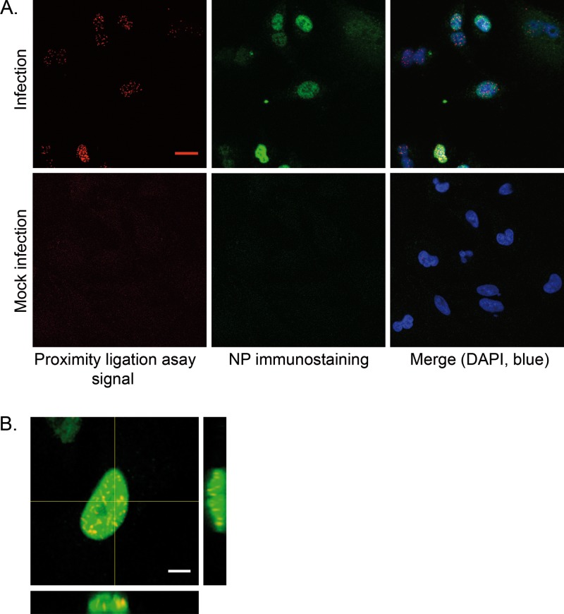
Association of endogenous HMGB1 with influenza virus NP in infected cells, detected in a Duolink II proximity ligation assay (PLA). A549 cells were infected with the WSN virus at a MOI of 3 PFU/cell (A, upper panels, and B) or mock infected (A, lower panels). At 4 hpi, the cells were subjected to Duolink II PLA as described in Materials and Methods, using primary anti-HMGB1 and anti-NP antibodies. In panel A, the PLA-specific fluorescent signal, the NP-immunostaining signal, and the DAPI nucleic staining were pseudo-colored red, green, and blue, respectively (scale bar, 20 μm). In panel B, orthogonal view of a three-dimensional reconstruction of an infected cell is shown, with PLA signal (red), NP immunostaining (green), and merge (yellow). Scale bar, 5 μm.

