
Fig 3.
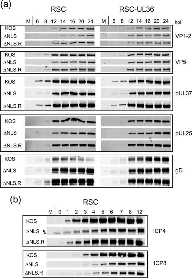
Comparison of protein expression during infection by K.VP1-2ΔNLS and its revertant. (a) RSC or RSC-HAUL36 cells were infected in parallel with the mutant virus K.VP1-2ΔNLS, the revertant virus K.VP1-2ΔNLS.R, or wt KOS virus at an MOI of 5. Samples were harvested at the indicated times, and expression of a series of delayed early or late proteins, i.e., VP5, pUL37, UL25, gD, and VP1-2 itself, was examined by Western blotting using fluorescent antibodies and quantitative detection as described in Materials and Methods. (b) Same as panel a, but examining immediate early (ICP4) and delayed early (ICP8) proteins every hour for 12 h. Some reduction in the abundance of ICP4 (long arrow) was observed, though there was little difference in the amount of ICP8. (A nonspecific band migrating just below ICP4 was occasionally observed, as indicated by the short arrow.) The parental KOS strain exhibited slightly increased expression of both proteins, estimated by quantitative analysis as a 2- to 4-fold increase over expression by the ΔNLS and ΔNLS.R viruses.