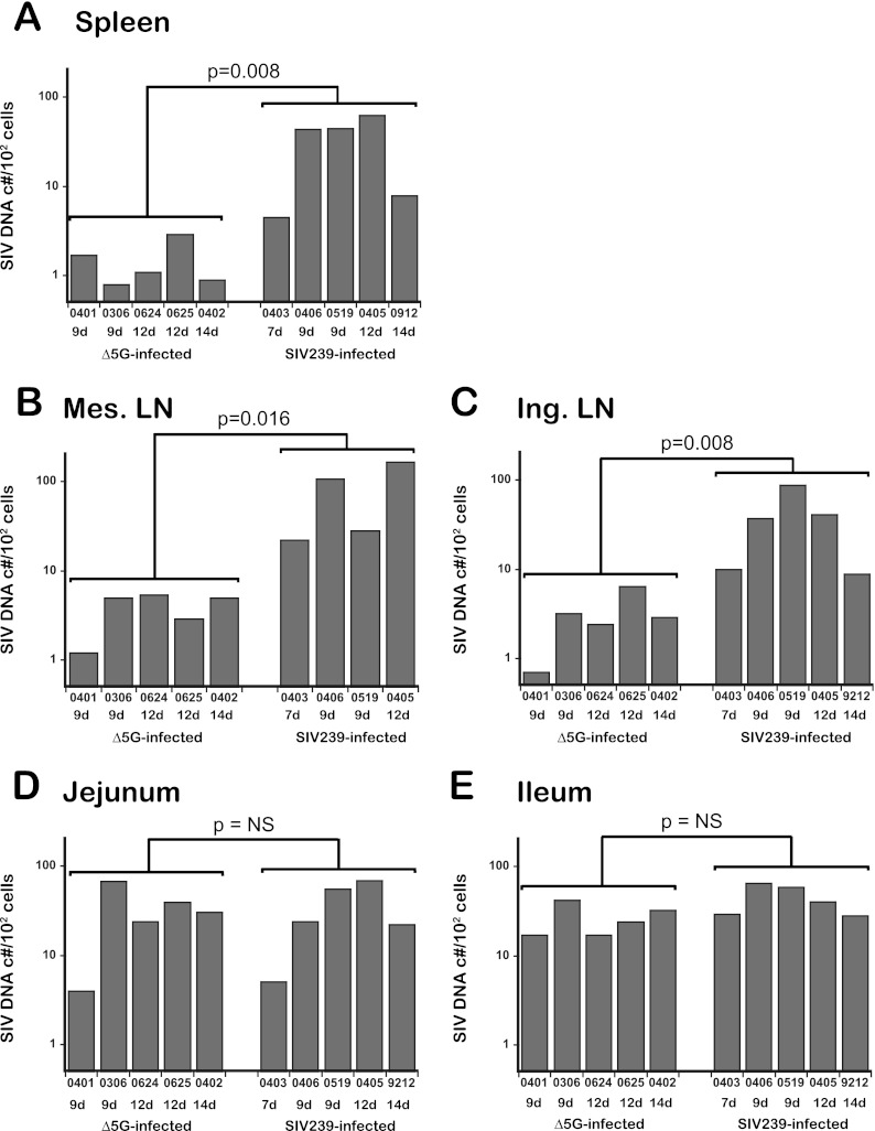
Fig 3.
SIV DNA loads in CD4+ T cells from intestinal and secondary lymphoid tissues. SIV DNA levels (SIV DNA copies/102 cells) in CD4+ T cells from the spleen (A), mesenteric LN (B), and inguinal LN (C) as representative secondary lymphoid tissues and from intestinal ileal (D) and jejunal (E) tissues from Δ5G- and SIVmac239-infected animals were determined by real-time PCR. The significance of differences in SIV DNA levels in CD4+ T cells between Δ5G- and SIVmac239-infected animals during the primary infection (7 to 14 days p.i. for SIVmac239-infected animals and 9 to 14 days p.i. for Δ5G-infected animals) was determined utilizing the unpaired Student t test. Differences were considered statistically significant when P values were <0.05. NS, not significant.

