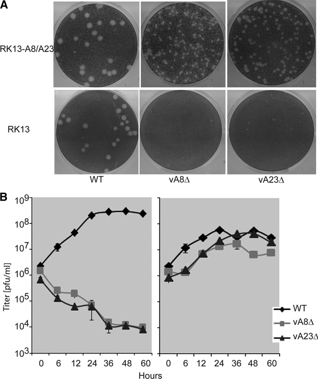
Fig 2.
Replication of A8R and A23R gene deletion mutant viruses in the complementing RK13-A8/A23 cell line. (A) Plaque formation. Suitable dilutions of WT VACV and mutant vA8Δ and vA23Δ VACV were added to RK13 and RK13-A8/A23 cell (clone number 11) monolayers and covered with a methylcellulose overlay. After 48 h at 37°C, the cells were stained with crystal violet. Plaques appear as round white areas against a dark background. (B) One-step growth curve. RK13 cells (left panel) or RK13-A8/A23 cells (right panel) were infected with 10 PFU/cell WT VACV or mutant vA8Δ or vA23Δ virus. The cells were harvested at the indicated times, and the virus titers were determined by plaque assay.

