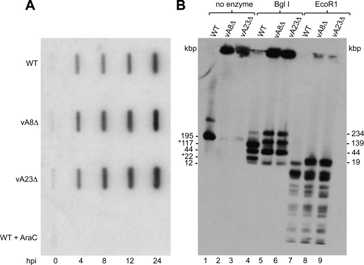
Fig 6.
Viral DNA synthesis and concatemer resolution. (A) Slot blot analysis. Total DNA samples isolated at 0, 4, 8, 12, and 24 hpi from RK13 and RK13-A8/A23 cells infected with WT, vA8Δ, or vA23Δ VACV or WT VACV in the presence of AraC were subjected to slot blot analysis using a digoxigenin-dUTP-labeled VACV DNA probe. (B) DNA from RK13 cells infected with WT, vA8Δ, or vA23Δ VACV was embedded in molten agarose, deproteinized, and incubated without restriction endonuclease (lanes 1 to 3) or with the restriction endonucleases BglI (lanes 4 to 6) and EcoRI (lanes 7 to 9). Following pulsed-field electrophoresis, the DNA was transferred to a membrane and hybridized to a digoxigenin-labeled VACV DNA probe. The sizes in kbp of full-length VACV genomic DNA and BglI restriction endonuclease fragments are shown on the left. Asterisks indicate end fragments. The sizes of the BglI and EcoRI concatemer junction fragments are shown on the right.

