Fig 4.
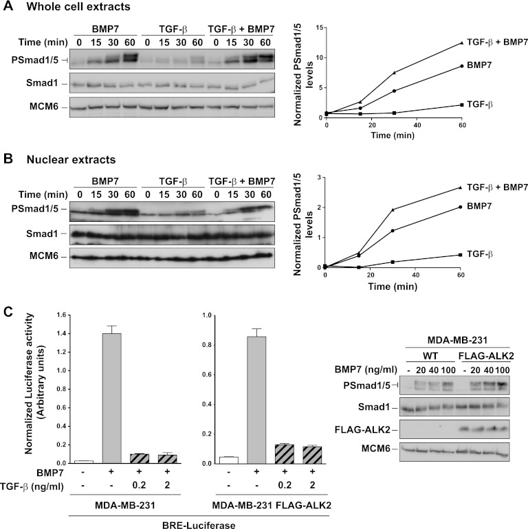
TGF-β does not inhibit BMP's ability to induce phosphorylation of Smad1/5. (A) Whole-cell extracts were prepared from MDA-MB-231 cells treated with BMP7 and/or TGF-β for the times shown. Extracts were Western blotted using antibodies against phosphorylated Smad1/5 (PSmad1/5), Smad1, and MCM6 as a loading control. The blot assays were quantitated on an ImageQuant LAS4000 mini, and the levels of PSmad1/5 relative to Smad1 are plotted on the right. (B) Same as panel A, except that nuclear extracts were assayed. (C, left side) MDA-MB-231 cells or the same cells stably expressing FLAG-ALK2 were transiently transfected with BRE-luciferase and TK-Renilla. After 24 h, cells were induced with the ligands as indicated for 8 h. Luciferase/Renilla assays were performed as in the legend to Fig. 1. (C, right side) Whole-cell extracts were prepared from the same cell lines induced for 1 h with different concentrations of BMP7. The extracts were Western blotted using antibodies against PSmad1/5, Smad1, FLAG, and MCM6 as a loading control. WT, wild type.

