Fig 9.
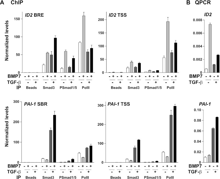
Smad3 and PSmad1/5 accumulate on BMP-responsive elements in response to costimulation with TGF-β and BMP7 in vivo. (A) ChIP analysis of the BRE and TSS regions of the ID2 gene and the SBR and TSS of the PAI-1 gene using antibodies against Smad3, PSmad1/5, and RNA PolII. Cells were starved overnight in Opti-MEM and then stimulated with the ligands indicated. (B) Cells were treated with the ligands shown for 1 h and analyzed by qPCR for levels of ID2 and PAI-1 mRNAs. In panels A and B, the data are means and standard deviations of qPCRs performed in triplicate in a representative experiment. To increase the sensitivity of these assays, we used a HaCaT cell line that stably expresses low levels of HA-Smad3 (data not shown).

