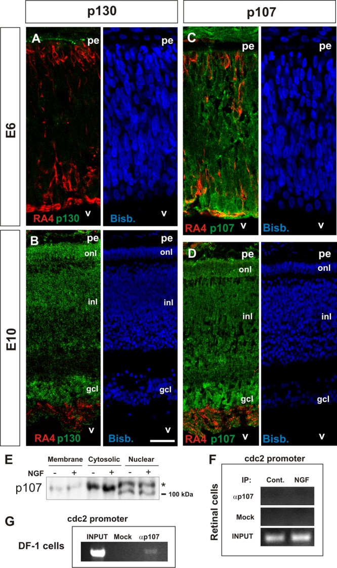
Fig 5.

Analysis of p130 and p107 expression in DCRNs and lack of interaction of the latter with the cdc2 promoter in these cells. E6 (A and C) or E10 (B and D) chick retina cryosections (12 μm) immunolabeled for either p130 (A and B) or p107 (C and D) (green) and the RGC-specific marker RA4 (red) and counterstained with bisbenzimide (blue). (E) Western blot with the anti-p107 antibody in the indicated subcellular fractions from DCRNs cultured for 30 min either in the presence (+) or absence (−) of NGF. The asterisk indicates the hyperphosphorylated 107. (F and G) ChIP analysis of the occupancy of the chick cdc2 promoter by p107 in either DCRNs cultured for 30 min either in the presence (NGF) or absence (Control) of NGF (F) or in DF-1 chicken fibroblasts (DF-1 cells) (G). Anti-p107 (αp107) or irrelevant (Mock) antibodies were used for immunoprecipitation. INPUT, RT-PCR amplification from extracts before immunoprecipitation. pe, pigment epithelium; v, vitreus body; gcl, ganglion cell layer; inl, inner nuclear layer; onl, outer nuclear layer. Bars: 30 μm (A and C) and 60 μm (B and D).
