Fig 2.
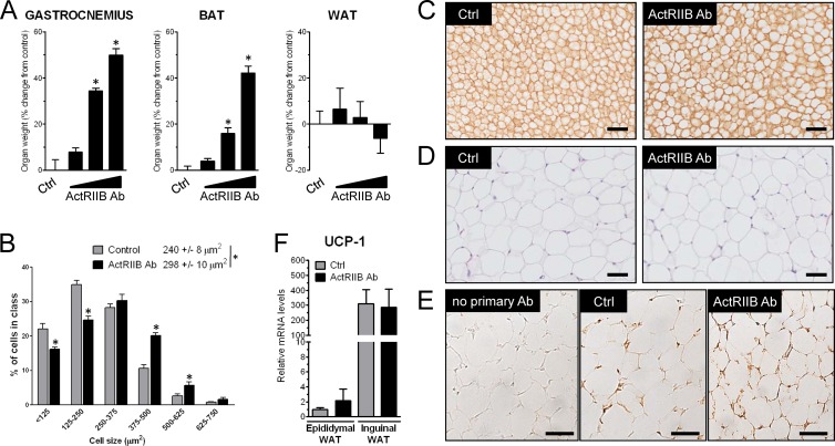
ActRIIB inhibition in mice increases the amount of brown but not white fat. (A) SCID mice (n = 12/group) were treated for 4 weeks with a weekly subcutaneous injection of control (Ctrl) antibody (IgG1; 20 mg/kg/week) or increasing amounts of a human monoclonal antibody against ActRIIB (2, 6, and 20 mg/kg/week). The wet mass of gastrocnemius muscle, interscapular BAT, and epididymal white adipose tissue (WAT) was measured and expressed as the percent change from the average of the control group. *, P < 0.05. (B and C) Interscapular brown adipose tissue from control and ActRIIB Ab-treated mice (20 mg/kg/week) was imaged by light microscopy after laminin staining, and cell size was quantified by histomorphometry. (D) Epididymal white adipose tissue from control and ActRIIB Ab-treated mice (20 mg/kg/week) was imaged by light microscopy after hematoxylin-eosin staining. (E) Inguinal white adipose tissue from control and ActRIIB Ab-treated mice (20 mg/kg/week) was imaged by light microscopy after UCP-1 immunohistochemistry. Bars, 50 μm. (F) Quantitative reverse transcription-PCR measurement of relative UCP-1 levels in epididymal and inguinal white adipose tissue from control and ActRIIB Ab-treated mice (20 mg/kg/week).

