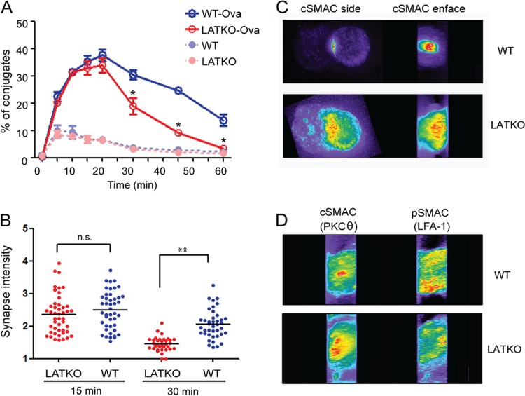
Fig 4.
Formation of CTL-target cell conjugates and the immunological synapse in LATKO CTLs. (A) Conjugate formation. CTLs were labeled with Cell Tracker Violet and mixed at a ratio of 1:1 with L1210/Kb cells labeled with Cell Tracker Orange with or without Ova peptide. At various time points, the cells were fixed and analyzed by FACS. CTL-target cell conjugates were defined as cells labeled with both fluorescent dyes. The graph shows one representative of three independent experiments. *, P < 0.05. Error bars represent means ± the SEM. (B) Quantitative analysis of cSMAC accumulation at the synapse of WT and LATKO CTLs. CTLs were incubated with L1210/Kb cells preloaded with the Ova peptide for 15 and 30 min to allow for the formation of conjugates. The conjugates were fixed, permeabilized, and stained with anti-PKCθ antibody to represent cSMAC and anti-LFA1 antibody to represent pSMAC. cSMAC accumulation was calculated as the ratio of the synaptic fluorescence intensity to the cytoplasmic fluorescence intensity. Each dot represents one CTL. Horizontal bars indicate the mean. n.s., not significant; **, P < 0.005. The data shown are from two independent experiments. (C and D) Immunological synapse patterns. The representative cells depicted here show the side and enface of cSMAC (C) and excluded pSMAC (D) of WT and LATKO CTLs.

