Fig 1.
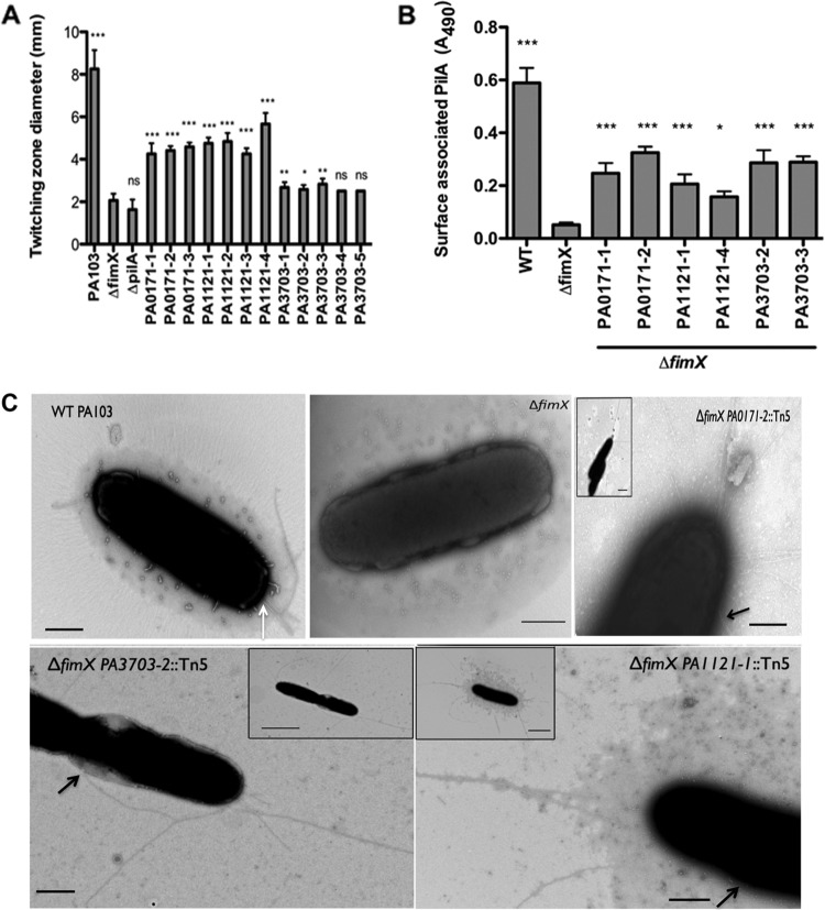
Extragenic suppressors restore surface pili. (A) Twitching zone diameter as measured by the subsurface stab assay. Strains were assayed 3 to 5 times, and means ± standard deviations (SD) (n = 4) from a representative experiment are shown. All of the strains were compared to ΔfimX using one-way ANOVA followed by Dunnett's multiple-comparison test. (B) Total surface pili as determined by surface pilin ELISA. Two representative Tn insertions were assayed for each gene. Bars indicate means ± SD from three independent experiments. All of the samples were compared to ΔfimX using one-way ANOVA followed by Dunnett's multiple-comparison test. *, P < 0.05; **, P < 0.01; ***, P < 0.001. (C) Visualization of T4P by transmission electron microscopy. Bacteria from early exponential growth phase were stained directly with 1% phosphotungstate. Panels show higher magnification pictures of representative cells for each strain, and insets show lower magnification pictures of the same cells. Black arrows indicate the nonpolar site of attachment of the pili with the cell body. White arrow indicates T4P on a WT surface. Scale bar, 500 nm (main panel) or 2 μm (inset).

