Fig 1.
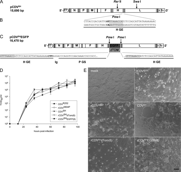
Construction and characterization of recombinant CDVs. (A) Schematic representation of pCDVSH full-length infectious clone. A subclone containing the H-L gene boundary was produced following digestion of pCDVSH with RsrII and SwaI to facilitate the introduction of a unique PmeI site (B) as indicated by the gray shading. (C) An additional transcription unit containing EGFP or dTom was inserted at the H-L gene boundary using the unique PmeI site to produce the pCDVSHEGFP(6) and pCDVSHdTom(6) full-length clones. (D) Multistep growth curves of CDVR252, CDV5804P, CDVSH, rCDVSHdTom(6), and rCDVSHEGFP(6) in VerodogSLAM cells. Virus was harvested at 12-h intervals up to 96 h postinfection. Viral titers were determined as the number of TCID50/ml in an endpoint titration test. Measurements shown are averages of triplicates ± SE. (E) Phase-contrast photomicrographs taken at 72 h.p.i. of VerodogSLAM cells with Opti-MEM (mock), CDVR252, CDV5804P, CDVSH, rCDVSHdTom(6), and rCDVSHEGFP(6). Bar, 200 μm.

