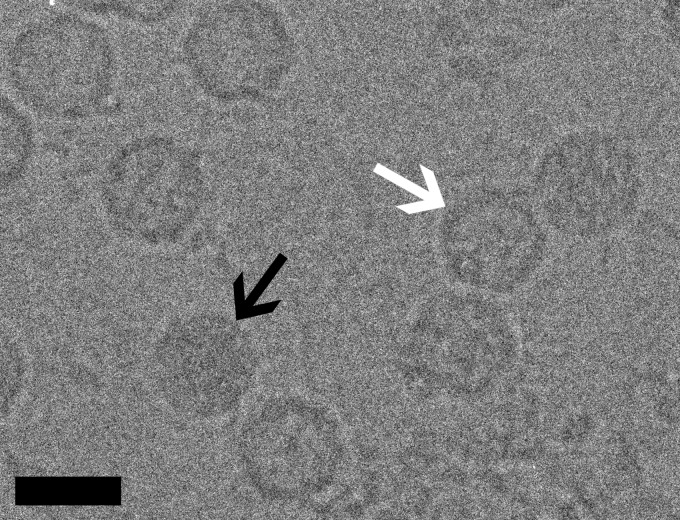
Fig 1.
Micrographs of CAV7 particles suspended in a layer of vitreous water over holes in a carbon support film were recorded at a nominal magnification of ×62,000 in an FEI Tecnai F20 microscope operated at an accelerating voltage of 200 kV. Black and white arrows indicate filled particles and empty capsids, respectively. This image was recorded at a 4.3-μm underfocus. Bar, 30 nm.

