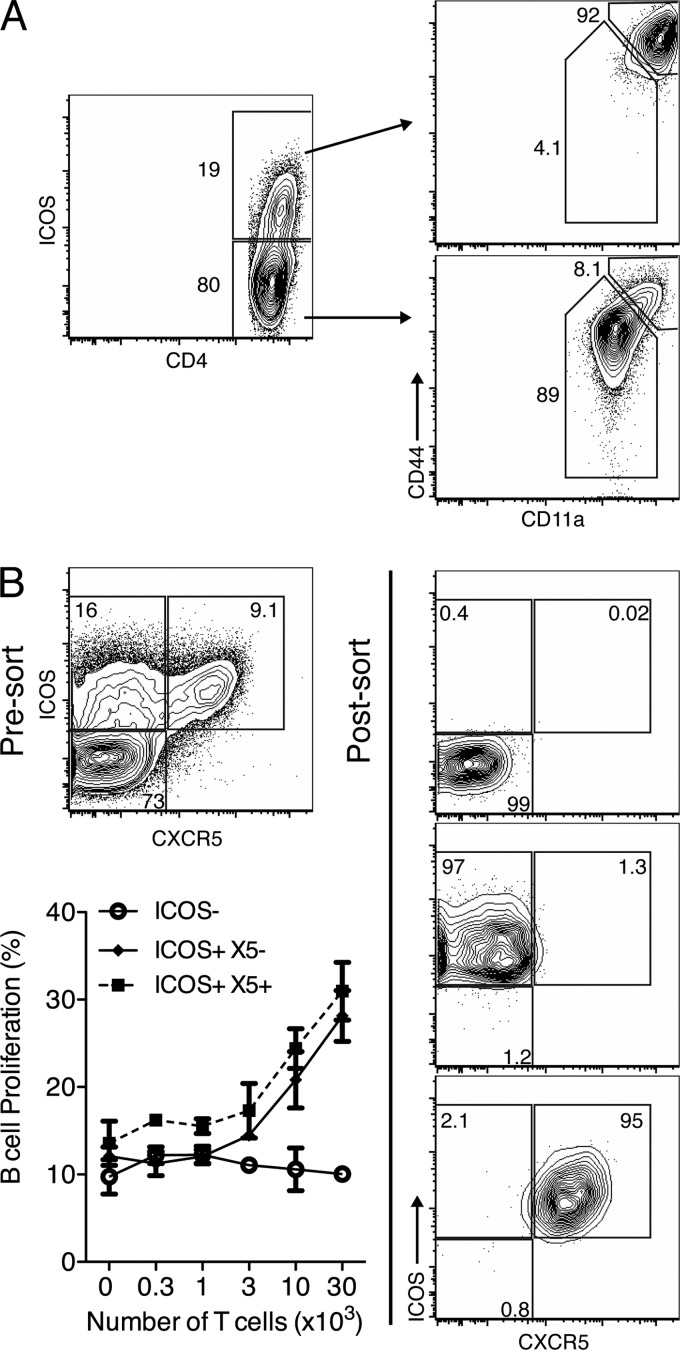
Fig 1.
B helper function is restricted to ICOS+ CD4 helper T cells. (A) BALB/c mice (n = 2) were infected with influenza virus A/Mem71 for 5 days and examined by flow cytometry. Shown are 5% contour plots with outliers gated on live CD3+ CD4+ lymphocytes, with percentages of CD3+ CD4+ cells or ICOS+ cells indicated. Plots are representative of data from 3 similar experiments using BALB/c, BALB/cByJ, or C57BL/6 mice. (B) BALB/c mice (n = 12) were infected with influenza virus A/Mem71 for 12 days, and CD4+ T cells were sorted from pooled MedLN based on ICOS and CXCR5 expressions. Shown are 5% contour plots with outliers of presort and postsort samples. Graded numbers of sorted T cells were cultured for 4 days with 2.5 × 10⁵ CFSE-labeled B cells from pooled inguinal lymph nodes (IngLN) of BALB/c mice (n = 4) at 12 days post-influenza virus immunization. Shown are mean frequencies ± standard deviations (SD) for live CD45R+ B cells which had proliferated (CFSElo) (left), from one representative experiment of two performed.

