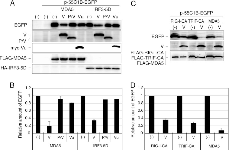
Fig 6.
IRF3 reporter assay in the presence of V proteins. (A) 293T cells were transfected with p-55C1B-EGFP together with pCAG-FL-MDA5 or pCAG-IRF3-5D and a plasmid expressing the V, P/V, or myc-Vu protein, as indicated. After 24 h, cells were solubilized and processed for Western blotting using anti-GFP antibody (EGFP), anti-P antibody (V, P/V), anti-myc antibody (myc-Vu), anti-FLAG antibody (FLAG-MDA5), or anti-HA antibody (HA-IRF3-5D). (B) The density of EGFP bands was quantitated and plotted in a graph. The value is the ratio against that without V protein [(−)]. Experiments were repeated at least three times, and the average value is shown. Error bars represent standard deviation. (C and D) 293T cells were transfected with p-55C1B-EGFP together with pCAG-FL-RIG-I-CA, pCAG-FL-TRIF-CA, or pCAG-FL-MDA5 and a plasmid expressing the V protein. After 24 h, cells were solubilized and processed for Western blotting, and the density of EGFP bands was quantitated and plotted in a graph as was done for panel B.

